
2013
Project types: GH indicates Global Health | MT indicates MedTech
Graduate
-
The Team
Student Team: Kaitlyn Harfmann, Ting-Yu Lai, Adam Lightman, David Narrow & Dr. Devin Coon
Clinical Advisers: Seth Goldstein and Ying-Wei Lum JHU SOM, Department of Surgery; Gedge Rosson, JHU SOM, Department of Plastic Surgery; Eduardo Rodriguez, University of Maryland/R. Adams Cowley Shock Trauma Center, Division of Plastic Surgery; Emad Boctor, JHU SOM, Department of Radiology; Jerry Prince, JHU Whiting, Department of Electrical and Computer Engineering
Technical Advisers: Drs. Soumyadipta Acharya, Emad Boctor, Jerry Prince & Youseph Yazdi
Abstract
Each year 50,000 patients in the U.S. alone will undergo free flap reconstruction, about 15% of whom will experience non-preventable vascular complications. These surgeries have the power to restore patients’ lives, yet too often they result in failure. If signs of a vascular flow problem are detected early, surgeons can repair the vessels before it is too late − thus, monitoring is pivotal to the success of these cases. However, half of these patients suffer from costly and morbid total flap failure due to inherent flaws in current monitoring devices, despite the fact that hospitals are paying hundreds to thousands of dollars to monitor each patient after surgery.
EchoSure has developed a novel monitoring technology with the power to prevent thousands of surgical failures and avoid unnecessary and expensive re-operations, while saving hospitals over $2,200 per surgery. Current competitors have high false positive rates, non-definitive outputs, and significant time delays to identify a problem; these factors lead to decreased surgical success, unnecessary procedures, and clinical frustrations. Our unique, clinically driven insight was that Doppler ultrasound was the ideal technology for monitoring — if it could be de-skilled so front-line clinicians could use it. The result was EchoSure, a dual-component system comprised of EchoMark and EchoFind. EchoMark is an implantable, resorbable marker placed beneath the vessels of interest, which acts as a homing beacon for nurses tasked with monitoring post-operative patients. EchoFind, a software application, is used to help guide users to the ideal position with the ultrasound at which point critical information about the vessel health is gathered and used to assess the patient. This technology will help save hospitals over $180M annually while decreasing unnecessary procedures and improving outcomes.
-
The Team
Sponsor: Jhpeigo and US AID/Saving Lives at Birth
Student Team: Dana Schultz, Hina Shah, James Su, Michelle Zwernemann, Vaishakhi Mayya
Abstract
Moderate to severe anemia is a major public health concern, particularly dangerous during pregnancy for both the mother and the baby. 100,000 maternal deaths and 600,000 neonatal deaths worldwide are attributable to anemia each year. The prevalence of anemia in the developing world is staggering, with estimates by the WHO of up to 50% among pregnant women and 65% among children.
There is a need for a device that can be used to widely and cheaply to screen for anemia. This device should enable identification of those at the highest risk, especially those with moderate-severe anemia in late gestation, in order to bring them “out of the cold” and into existing healthcare structures. Moreover, a device that is able to track patient data geographically and over time would facilitate macro-scale public health policy by enabling the targeting of health care initiatives to areas in need and by providing feedback on interventions. Such a device would have to be non-invasive and affordable.
Hemoglobe is a non-invasive anemia screening device that leverages the existing cell phone technology to provide a cost effective method for screening for anemia, while providing real time surveillance data about the status of the pregnant woman.
-
The Team
Student team: Ting-Yu Lai, Adam Lightman, David Shin, and QX Yee
Clinical Advisors: John Sampson, MD, Department of Anesthesiology and Critical Care Medicine; Dr. Lynn Kanyuuru, Jhpiego Kenya and Dr. Kusum Thapa, Jhpiego Nepal
Sponsor: Jhpiego
Abstract
Oxygen is a critical, life saving medicine. It is used to manage childhood pneumonia, COPD, obstetric emergencies and is necessary for performing surgeries. Despite these facts, oxygen is generally not available below the district hospital level in the developing world. There are multiple technologies available for supplying patients with oxygen in the developing world, yet no technology has proven to be a sustainable solution for resource starved clinics.
Our team has analyzed oxygen delivery from the perspectives of technological solutions, business model innovations, and policy level efforts. Using this knowledge, we have devised a sustainable business model to improve the availability of concentrated oxygen in lower-level facilities in developing countries. In this model, local entrepreneurs purchase kits of affordable oxygen concentrators and bottling equipment and sell filled oxygen cylinders to health facilities within a catchment area. The model incentivizes equipment maintenance and simplifies distribution, common pitfalls of previous attempts to provide concentrated oxygen to lower-level facilities. When combined with a parallel effort by NGOs to promote oxygen demand, we project our model to create a sustainable enterprise while dramatically increasing the availability of oxygen.
-
The Team
Student Team: Stephen Dria, Kaitlyn Harfmann, Christopher Lee, David Narrow
Clinical Advisors: Janine Bullard, MD, Department of Neonatology, JHU
Other Advisors: Helge Myklebust, Harshad Sanghvi, MD
Sponsors: Day of Birth Alliance (CBID, Jhpiego & Laerdal Global Health)
Abstract
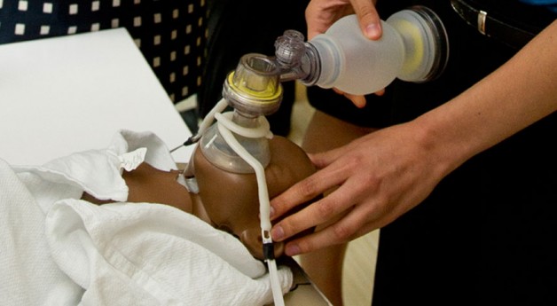
Annually, there are over 4 million global neonatal deaths primarily occurring in resource-constrained environments. This accounts for over 38% of deaths in children under the age of five. When performed properly, neonatal resuscitation is an effective means to revive newborns that are unable to properly breathe. Statistics indicate that birth asphyxia accounts for 23% of all neonatal deaths. Additionally, another one million newborns suffer from permanent disabilities due to insufficient oxygen at time of birth. Effective newborn resuscitation alone can prevent millions of newborn deaths. Neonatal resuscitation requires the use of a ‘bag-valve-mask’ or BVM. The BVM channels ambient air from the atmosphere into a one-way valve, when the user compresses the bag-component. Gas is then expelled through the mask and into the trachea, bronchus and lungs of the infant. Due to the technical difficulties associated with the procedure, newborns that otherwise would have been resuscitated die because of ineffective equipment operation and improper technique—a function of the burden of training and skill retention. Thus, it is critical to reduce the amount of skill required for providing an effective and consistent resuscitation, especially in peripheral health care centers of the developing world.
Failed resuscitations are attributed to poor head positioning and an inadequate seal between the mask and the baby’s mouth. However, air is forcibly pushed into the lungs through ventilation only ;when proper mask seal and head positioning are addressed together. After significant research, chest rise is a variable that is most cost-effective to quantify successful resuscitation instantaneously. Thus, a tool to quantify lung expansion would be revolutionary to the resuscitation protocol, providing low-skilled health workers real-time feedback throughout the procedure. Our innovation will mitigate existing inadequacies attributed to constrained resources, diminished skill retention over time and difficulty using devices associated with resuscitation. Our technical solution quantifies chest expansion and guides the user through a proper resuscitation, improving the results of attempted resuscitations and successfully managing birth asphyxia.
-
The Team
Student Team: Christopher Lee, Dana Schultz, David Shin & Michelle Zwernemann
Clinical Advisors: Alan Schwartz, MD, Philip L. Smith, MD, Jason Kirkness, PhD and Hartmut Schneider, MD, PhD (Johns Hopkins Sleep Disorders Center)
Other Advisors: Susheel Patil, MD, PhD, JHU Sleep Disorders Center, Soumyadipta Acharya, MD, PhD
Abstract
39 million Americans suffer from sleep apnea (SA), a serious medical condition that causes people to periodically stop breathing during sleep. Established medical research has conclusively linked SA to many downstream clinical diseases such as heart disease, stroke, and cognitive atrophy. Economic analysts have estimated undiagnosed and untreated SA has an annual cost of $45–80 billion in medical costs and up to $165 billion in total downstream economic costs in the United States. Despite these serious consequences, approximately 2 out of 3 Americans with SA remain undiagnosed. SA diagnosis requires an overnight sleep study in a fully equipped and staffed sleep center. These studies comprehensively diagnose SA by monitoring up to 10 modes of physiologic data. However, this is not only expensive and uncomfortable for patients, but current sleep clinic infrastructure is unable to meet high patient demand. As a result, 26 million Americans suffering with SA remain undiagnosed.
To overcome the diagnostic bottleneck imposed by limited clinic test infrastructure, at-home sleep monitors have been introduced that allow patients to be screened from their own bedrooms. However, no home system exists that can be self-applied by patients to record all 10 necessary physiologic signals needed for a comprehensive diagnosis. As a result, home monitors have gained limited clinical adoption and represent only 2.5%. Our team is developing The Lyra, the first sleep apnea diagnosis system that effectively transitions sleep apnea diagnosis to the home setting. The Lyra integrates traditional sleep diagnostic technology and novel dry electrodes into comfortable, sleep-friendly form factors, a headset and chest strap, which record a complete dataset equivalent to an in-clinic sleep study.
-
The Team
Sponsors: Center for Tuberculosis Research Laboratory & Jhpiego
Clinical Advisors: Gyanu Lammichane, PhD & Yukari Manabe, MD – Center for TB Research; Stacie Stender, RN – Jhpiego
Student Team: Anjana Sinha, Hiren Mistry, Anmol Chopra, Hector Neira, Devin O’Brien-Coon
Abstract
Globally, only 60,000 of the estimated 310,000 new cases of Multi-Drug Resistant Tuberculosis (MDR- TB) are diagnosed. The situation is even more dismal in the 27 nations that account for 80% of MDR-TB cases, where less than 5% of patients receive an official diagnosis. MDR-TB diagnostics are typically available only at reference level facilities, two or three level above where patients actually seek treatment. Clinicians are therefore forced to “treat” these patients with highly ineffective drugs for at least six months, prior to modifying treatment. In the meantime, patients are likely to become increasingly resistant to treatment, and able to spread this airborne infection within their community. Progression of MDR-TB cases into extensively drug resistant (XDR-TB) cases is a major public health concern because XDR-TB strains are virtually untreatable. Our team is developing a self-contained assay comprising effective, yet inexpensive components, to bring the gold standard of care, culture-based diagnosis, to rural healthcare facilities. User-centric design is being employed to assure the final system enables minimally trained personnel to conduct MDR-TB testing safely, without sophisticated laboratory equipment. Expanding availability of MDR-TB diagnostics to rural health facilities will lead to adequate treatment for patients, while they are still responsive to available drugs.
-
The Team
Student Team: Anjana Sinha, Hector Neira, Qing Xiang Yee, Vaishakhi Mayya
Clinical Advisors: James Shin and Ashley Cimino-Mathews, MD. Dept of Pathology Melissa Suzzane Camp, MD MPH, Johns Hopkins Breast Center
Abstract
Patients undergoing Breast Conserving Surgery (BCS) have a one in five chance of requiring a reoperation because surgeons cannot determine if the entire tumor has been removed. Surgeons rely on pathologists to assess margins through histological evaluation. Due to the inherent mechanical properties of breast tissue, this cannot be done before the surgery ends. ClearView allows pathologists to produce high-quality histology slides within minutes, enabling intra-operative assessment of tumor margins. This could prevent up to 66,000 unnecessary BCS procedures in the U.S. annually.
Using ClearView, a typical surgery will require $120 of disposables, covered by insurance companies under existing reimbursement codes. According to FDA regulations, ClearView is a Class I device, the lowest-risk device category. This affords a quick time to market, with minimal testing and regulatory requirements. Our team is collaborating closely with leading clinicians from the Johns Hopkins Breast Center, who are excited about our initial results.
-
The Team
Student Team: Anmol Chopra, Hiren Mistry, Michael Batista, Hina Shah, James Su, Stephen Dria
Advisors: Inder Makin MD PhD, John Sperati MD, Robert Fitzgerald MD, Youseph Yazdi Ph.D MBA.,Ying-Wei Lum MD
Abstract
Hypertension, or high blood pressure, affects one-third of all Americans, increases the risk for heart disease, stroke and renal disease, and is directly associated with 350,000 American deaths annually. In the US, the annual cost attributable to hypertension is $250 billion in medical expenses. Currently, antihypertensive drugs are the only treatment. However, these therapies fail for 20% of hypertensive Americans that cannot control their blood pressure even when using multiple antihypertensive medications. These patients have a condition known as resistant hypertension. Two experimental device-based treatments have shown promise, but are currently in clinical trials: renal denervation (RDN) and baroreceptor stimulation (CBS). However, RDN excludes one third of patients and may damage the renal artery. Meanwhile, CBS requires the invasive implantation of a device and has failed safety endpoints due to severe procedural complications. Therefore, there is a need for a novel device-based treatment to help the millions with resistant hypertension.
The proportion of hypertensive Americans continues to grow and the market for device-based therapies for hypertension continues to expand. SympSolutions has developed AccuRIGHT, a novel device to treat resistant hypertension within a clinician’s office. The device utilizes the well established technology of high intensity focused ultrasound to noninvasively eliminate the carotid body, a central contributor to hypertension, without an operating room. In past animal and human trials, surgical removal of the carotid body has proven both safe and effective, creating reductions in pressure sufficient to restore normal blood pressure values in patients. In terms of development, initial feasibility animal trials have begun, ultrasound simulations have been performed to evaluate initial safety profiles, and a therapeutic ultrasound setup has been proven to generate lesions sufficient to eliminate the carotid body.
As this novel device avoids the shortcomings of other device-based treatments, patients will see improved health outcomes, health care providers will gain a new source of revenue, and the cost burden to the healthcare system will be reduced.
Undergraduate
-
The Team
Student Team: Manjima Dhar, Yun-An Chen, Alex Dakos, W. James Melvin, Justine Yu, Jennifer Zheng, Tony Wu
Clinical Advisor: Elliott Schwartz, DDS
Abstract
Oral cancer has a 45% fatality rate within five years of diagnosis. This high fatality rate is due to late stage detection. The symptoms of oral cancer are lesions in the oral cavity. However these symptoms are not unique to oral cancer, therefore clinicians cannot easily distinguish cancerous lesions by visible inspection.
To help screen for oral cancer, we have designed and prototyped OralCheck: a portable cell analyzer. OralCheck has two components: a brush for sample collection from the mouth and a microfluidic chip for analysis. The brush is able to collect cells from the lower levels of the oral cavity epithelium where cancer arises. A syringe is attached to the inlet to allow easy sample insertion. The microfluidic analyzer uses electric fields to sort the cancer cells from benign cells into separate compartments. The sorted cells will react with a chemical to give a color change that is visible to the naked eye. A positive result will show two visible dots (one in each well), and a negative result will show only one. OralCheck gives the dentist a quick and easy way to check for cancer cells in any suspicious lesion.
-
The Team
Student Team: Jonathan LeMoel, Jay Bhasin, Rob Hubbard, Sean Reeder, Jimmy Su, Grant Kitchen, Inez Lam, Annie Mao
Clinical Advisor: Hien T. Nguyen, M.D., Department of Surgery, JHH
Abstract
In about 3,000 open surgeries each year, a surgical sponge is inadvertently left inside the patient, requiring a second surgery to remove it, at a cost of $700M to health care system. This is in spite of a “correct sponge count” by the nurses and OR technicians after every surgery. To reduce the number of these second operations, we have developed a wrist-mounted device worn by the circulating nurse integrating seamlessly into the existing workflow of the OR. Our device, Haptact, uses radio-frequency antennae affixed to the wrist of the nurse’s sterile sleeve to detect unique RF identification tags on the sponges. As the nurse picks up the sponge during the customary manual count, Haptact keeps track of that sponges unique tag number, preventing duplicate counts and keeping a running update of exactly what is “checked out” and in use and what has been “checked in” and returned after the surgery. Haptact thereby supplements the manual counting technique to reduce the risk of an incorrect count.
-
The Team
Student Team: Joshua Budman, Valeriya Aranovich, James Frick, BaDoi Phan, Paul Tershakovec, Doran Walsten, Ben Wheeler, Alp Yurter
Clinical Advisors: Jennifer Monti, MD
Abstract
p>Approximately 10 million Americans have PVD, and the disease affects almost 20 percent of the population over 65 years old. Smokers and diabetics are especially susceptible to the disease. The disease is a general term that describes obstructions of the large arteries excluding the coronary artery, the aortic arch and the brain. The disease is often characterized by swelling in various areas of the body, pain while walking short distances and in more serious cases pain at rest. When the disease is highly evolved in certain individuals, gangrene may develop.
Pre-surgical treatments to PVD include various drug regimens, including blood-pressure medication and cholesterol medication, which are highly expensive and are accompanied by a score of life-altering side effects. Additionally, lifestyle changes such as dieting, exercise and cessation of smoking, while effective, are very difficult to enforce by physicians. The Human-Ultrasonic Mesenchyme-Stimulating (HUMS) Device relies on the application of high frequency stimulation, in the ultrasound frequency range, to areas with a high density of mesenchymal tissue in order to mimic the longitudinal stresses of exercise in these areas and release endothelial progenitor cells (EPCs) from the mesenchyme into the peripheral blood. Research has shown that an increased EPC count has a causal relationship with reduction of PVD symptoms. The peripheral application of the device makes it non-invasive, and since it is a one-time purchase it is more cost-effective than drugs. Finally, its easy application will make it a more appealing treatment than change of lifestyle.
-
The Team
Student Team: Rowan Cade, Rochelle Dumm, Ian Graham, Brian Jeon, Kevin Keenahan, Kevin Moon, German Om, Emily Rencsok
Clinical Advisors: Mark Hopkins, PT, CPO, MBA, PM&R, JHMI, Dr. Lew Schon, M.D. Union Memorial
Abstract
Two thousand victims, every month, are maimed by landmines worldwide and 80% of these patients are unable to receive suitable prosthetic legs. The ones fortunate enough to receive any device at all walk on poorly fit sockets that can be extremely painful. Prosthetic legs in the US and abroad are extremely expensive and, considering a patient can go through at many as 25 throughout his or her lifetime (due to limb atrophy and fit considerations), these costs can become insurmountable for patients living on less than a dollar a day.
We are developing a new system to solve these problems. We are designing a device that can be remolded to fit the patient’s changing residual limb. This reduces or eliminates the need for these costly replacement devices and could save the amputee thousands of dollars over a lifetime. In addition, we are decreasing fitting time from one week to only hours by eliminating the need for expensive and time-consuming mold making procedures. Our device can be molded directly to a patient’s limb, sized, and assembled in one visit. We are hopeful our device will be successfully implemented and will assuage the suffering of the nearly 10,000 new amputees every year who are denied access to appropriate prostheses.
-
The Team
Student Team: Anne Pigula, Malvi Hemani, Hyun Soo Jang, Yong Kim, Megan Lamberti, Angelica Herrera, Barbara Kim, Brian Gu
Sponsors: Laerdal Global Health and Jhpiego
Clinical Advisors: Sheena Currie, RM, Jhpiego
Abstract
Roughly one million newborns die every year due to difficulty breathing at birth, but many of these lives could have been saved by proper neonatal resuscitation. We have developed Neo2Inspire, a suite of tools to meet these gaps in resuscitation. First, we have developed a plastic mat which provides support while positioning the infant to achieve an open airway. This mat folds up into a box, which contains the bag-valve-mask resuscitator and other tools. Second, we have incorporated elastic straps to secure the mask onto the infant’s face, ensuring an airtight seal. Finally, we have implemented a heat-producing solution, which utilizes a reusable chemical packet to prevent infant hypothermia. Our solutions are inexpensive, easy to clean, and are supported by extensive end-user and laboratory testing. Neo2Inspire will help health care providers to bring life to every child.
-
The Team
Student Team: Vikram Rajan, John J. Kim, Connor Jacobs, Tony Wei, Daniel Levenson, Edric Tam, Ivan Kuznetsov, Monica Rex, Ravi Gaddipati
Clinical advisor: Dr. Michael V. Boland, M.D., Ph.D., Wilmer Eye Institute
Faculty advisor: Dr. Xingde Li, Ph.D., Department of Biomedical Engineering
Abstract
Glaucoma is the leading cause of irreversible vision loss. Astonishingly, up to 80% of afflicted individuals in developing world nations are unaware of their illnesses. To mitigate the impact of glaucoma, we have designed an affordable and accurate glaucoma screening device. Based on the principles of fundus imaging, this device outputs a structural image of the retina that functions as an indicator of the developing pathology of the disease. The novel features of our device are designed to make the learning curve for healthcare workers short. The device’s components are optimized to lower cost while not sacrificing intended screening-grade functionality. Costs and time are further minimized by segmented imaging and analysis hardware.
-
The Team
Student Team: Maher Khalil, Arjun Khakhar, Christina Jacob, Theodore Leclere, James Chuang, Jesse Zhang
Faculty Advisors: Dr. Marc Ostermeier, Dr. David Sullivan
Clinical Advisors: Dr. Robert Hamilton
Abstract
To help clinicians in the developing world test for the presence of malaria, we have designed a novel point-of-care diagnostic device. Our device focuses on an alternative method of diagnosis which will be economically feasible for developing countries. In addition, the device is suitable for implementation in areas with limited pre-existing healthcare infrastructure and limited training of healthcare workers.
Our device is a lateral flow assay that is able to detect malaria during the early stages of infection. Our novel platform replaces the antibodies that are generally used in such assays with bacteria expressing evolved scaffold proteins on their surface. Additionally, the device tests a patient’s urine for our biomarker which will obviate the need for skilled professionals to draw blood. Our test can rapidly and cheaply diagnose malaria so that patients can be diagnosed before treatment, preventing the spread of drug resistant malaria.
-
The Team
Student Team: Nick Gisolfi, Steven Albers, Anthony Alers, Margaret Chow, Ran Liu, Travis Wallace
Clinical Advisor: Ray Dorsey, MD, Department of Neurology
Abstract
We are developing a novel system for quantitatively and remotely assessing the severity of Parkinson’s disease (PD) symptoms. Since the mechanisms underlying PD are largely unknown, patients are treated symptomatically. Currently, a movement disorders specialist (MDS) administers a test which qualitatively evaluates the severity of symptoms associated with PD. The ultimate goal of our proposed system is to replace the qualitative nature of this test with a quantitative measurement which exhibits increased spatial resolution over the state of the art. Additionally, we aim to increase the temporal resolution. An MDS’s time is limited and traveling is burdensome to the patient, so we want to develop our system such that the remote assessment of a patient during their daily routines becomes a possibility. This will allow the construction of a database of patient specific data which an MDS can view in order to examine how a patient’s symptoms vary between successive doses, different activities, or subsequent assessments. With such a system, an MDS will have a much more descriptive image of a patient’s disease state than they would otherwise obtain through a routine office visit. This will allow an MDS to make more informed decisions when prescribing treatments, which will improve clinical outcome.
-
The Team
Student Team: Bijan Abar, Wes Bernier, Calvin Chang, Emily Hsiao, Arianne Papa, Sohini Sengupta, Jiarui Wang, Jennifer Yang and Amadeus Zhu
Clinical Advisors: James H. Black, M.D., FACS, James H. Black, M.D., FACS
Abstract
Trauma injuries to the inferior vena cava (IVC) are a serious, fatal matter with a patient mortality rate that exceeds seventy percent. These injuries are commonly caused by gunshot wounds, stab wounds, physical abuse, or rapid deceleration. Due to the high rate of blood flow through the IVC, it is imperative for the surgeon to stop the bleeding from the vein as quickly as possible. However, this is difficult to achieve in an open surgery setting due to the placement of the IVC within the body.
We have developed the ShunTube, an adjustable double balloon catheter that can be deployed without the need for open surgery. The device includes two occlusion balloons, located above and below the site of injury, that are both inflated with saline in order to seal off the wound. Blood will enter the device through pores located beneath the lower balloon, travel upward bypassing the wounded region, then exit above the upper balloon and return to the heart. Our device can be rapidly deployed to improve patient outcome and reduce medical expertise required for surgical intervention. Aside from treating trauma injuries, our device can also be modified for use in liver cancer treatment, deep vein thrombosis treatment, and as a substitute for veno-venous bypass in tumor resection procedures.
-
The Team
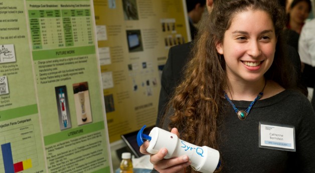
Student Team: Truc Nguyen, Daniel Adler, Catherine Bernstein, Alexia Haralambous, Jonathan Hunt, Thomas Nguyen, Antonio Spina, Lucia Tellez
Sponsor: Mitch Zhao, PhD, Janssen Research & Development
Abstract
In order to facilitate subcutaneous delivery of therapeutic monoclonal antibody (mAb) suspensions, we developed Syr-Q, an injection device. Syr-Q resuspends sedimented mAb particles that would otherwise clog the needle, leading to incomplete drug delivery. Syr-Q features an eccentric rotating mass (ERM), which adequately resuspends sedimented mAb particles by generating vibrations through centripetal acceleration. The ERM is attached to a timing circuit, which resuspends the particles for 15 seconds before shutting off, which allows the patient to safely self-administer the medication. Preliminary testing has shown that our resuspension mechanism is capable of decreasing the injection force from 80 N to less than 25 N, with statistical significance of p < 0.0005. Our proof of concept will allow for the self-administration of mAb therapeutics at home, saving on clinical resources and facilities, and will allow for the delivery of drugs that are currently undeliverable subcutaneously.
-
The Team
Student Team: Piyush Poddar, Aaron Chang, Melinda Chen, Peter Malamas, Sandya Subramanian, Joon Eoh, Kevin George, Rohil Malpani
Sponsor and Clinical Advisor: Todd Cohen MD
Abstract
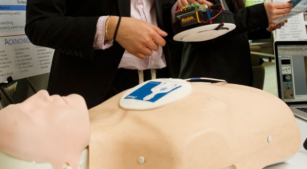
The PrestoPatch Integrated Electrode System allays the two major shortcomings of defibrillation and cardioversion: 1) Difficulty in switching the shocking vector between shocks 2) Difficulty in applying standardized external pressure to the patches to reduce transthoracic impedance. With regards to the shock vector, current patches are not meant to be relocated. Removing and re-applying the patches is difficult, time-consuming, and ineffective. Furthermore, applying an entirely new set of electrode patches is inefficient and not cost effective. For external pressure, literature has indicated that exerting firm pressure over the patches decreases transthoracic impedance. To accomplish this right now, clinicians use their fists or towels to press down on the patches. Not only is this method crude, but it also puts clinicians at risk of shocking themselves.
The complete PrestoPatch system solves these problems via three separate components:
- The Tri-Patch, a disposable three-patch electrode that allows for a instantaneously switchable shocking vector
- The SyncStrap, an elegant, yet disposable, device that reduces transthoracic impedance within 25 seconds
- The Sync Adaptor, a universal adaptor that makes the PrestoPatch system compatible with every defibrillator on the market today
This novel patch system will provide the ability to switch shock vectors quickly and with high reliability to offer physicians more versatility during shock therapy. It will also reduce the patient’s transthoracic impedance to more efficiently deliver the shock to the heart
-
The Team
Student Team: Tonia Wu, Garren Angacian, Ashley Cook, Gaby Frid, Woojin Kim, Jennifer Hui, Barry Leybovich, Molly Moore, James Verdone
Advisor: Dr. Edith Gurewitsch (Johns Hopkins Hospital Labor & Delivery
Abstract
Instrument-assisted vaginal delivery occurs in 7.4% of births in the United States; while it is safer than a cesarean section, instrument-assisted vaginal delivery requires significant skill and experience. Forceps delivery carries a 3.7 odds ratio of 3rd and 4th degree perineal lacerations over spontaneous vaginal delivery. These lacerations and stretching of the pelvic floor muscles can lead to increased risk of long-term complications with treatment amounting up to $40,000 over time. To assist obstetricians during forceps delivery, we plan to design and develop an instrument that will mitigate the risks of complications to the mother while safely delivering the fetus. The project aims to quantify the safe range of angle of traction and the point in which the fetal head clears the pubic symphysis during an instrument-assisted vaginal delivery. Our device will guide the physicians during the backward traction of the obstetric forceps by providing physicians with real-time feedback of angle of trajectory and position of the forceps in relative to the mother’s pubic bone. Efficacy will be demonstrated by a comparative study of the new device and classical forceps as applied to birth simulators. We aim to design a tool that will improve clinical techniques and clinical outcome significantly.